

What is Rihnolaryngoscope
A rhinolaryngoscopc, or nasal endoscope, is a medical instrument used to visualize the inside of the nose, throat, and larynx, aiding in the diagnosis and treatment of ENT(ear, nose, and throat) conditions.
Main Classifications
Video Rihnolarygoscope
Equipped with high-definition cameras, it transmits images to monitors for magnified observation, aiding in detecting subtle lesions. Superior in clarity compared to fiber optics but less flexible due to rigid construction.
Fiber Rihnolaryngoscope
Made of flexible fiber-optic material with a slender diameter, it offers maneuverability to examine nasal cavity corners. lmages are transmitted via optical fibers, though with relatively lower resolution.
Rigid Rihnolaryngoscope (Nasal Endoscope)
Constructed from hard materials with diameters of 2-3.5mm (thinner for pediatric use), it provides direct visualization of nasal and nasopharygeal areas.Offers stable imaging but may cause discomfort or tissue irritation.
Additionally, multifunctional devices like video rihnolaryngocopes allow combined examination of nasal and throat regions through a single insertion.
Selection depends on clinical needs (e.g. lesion screening, pediatric use) and patient tolerance.
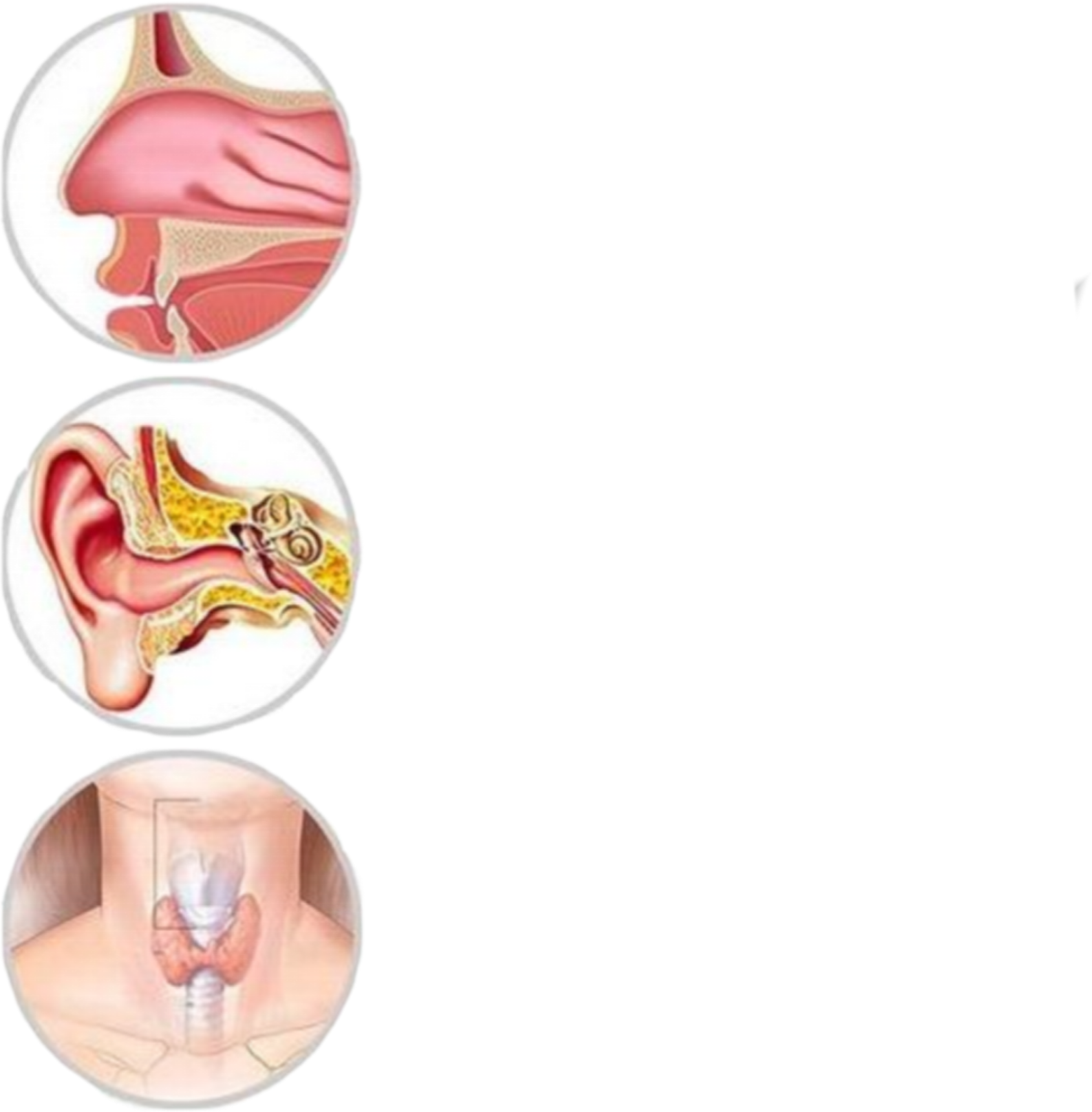
Indications
Rihnolaryngoscopy is primarily used to investigate and evaluate the following symptoms or clinical needs:
⚫ Nasal symptoms:
Recurrent nasalobstructon,rhinorrhea (runny nose), epistaxis (nosebleeds),blood-tinged postnasal drip, abnormal olfaction.
⚫ Throat symptoms:
Hoarseness, dysphagia (diffculty swallowing), throat foreign body sensaton,chronic cough.
⚫ Ear symptoms:
Unilateral or persistent secretory otitis media, tinnitus, hearing loss (potentially related to Eustachian tube dysfunction).
⚫ Other symptoms:
Sleep apnea, snorig, adenoid facies in children (e.g., chronic mouth brcathing).
⚫ Tumor screening:
Early detection of nasopharyngeal carcinoma in high-risk populations(e.g.,EBV infection, family history).
⚫ Postoperative assessment:
Monitorng recovery after nasal or nasopharyngeal surgery.
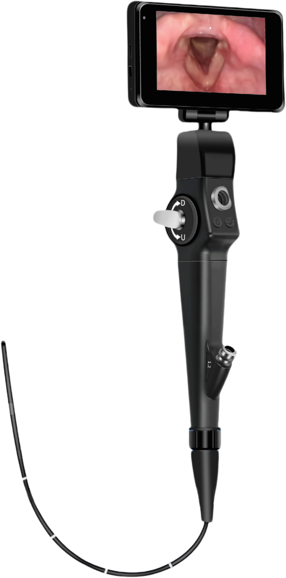
Features of MG-RI04
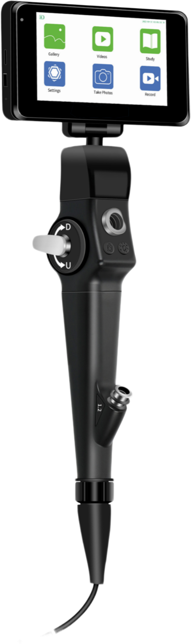
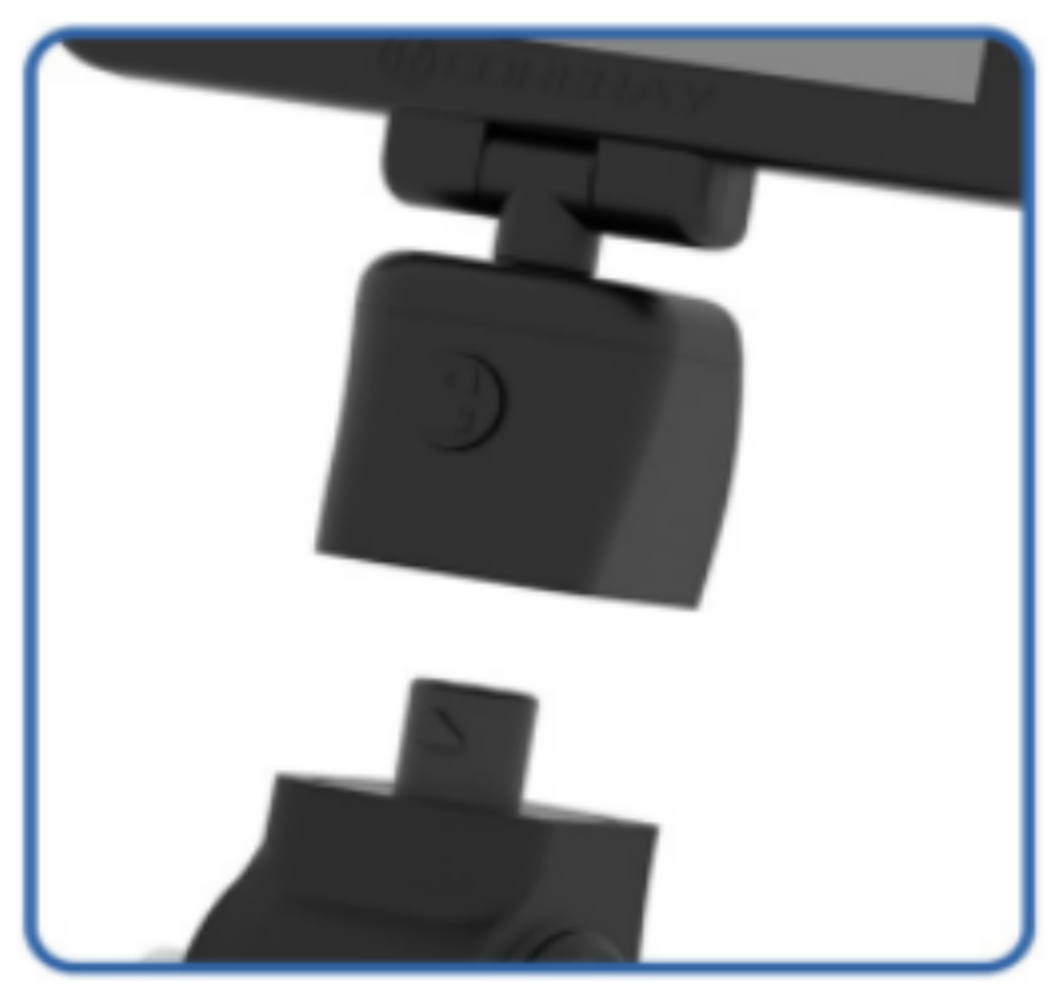 Quick Handlings
◾ 1 lock design
◾ Fast installation and disassembling
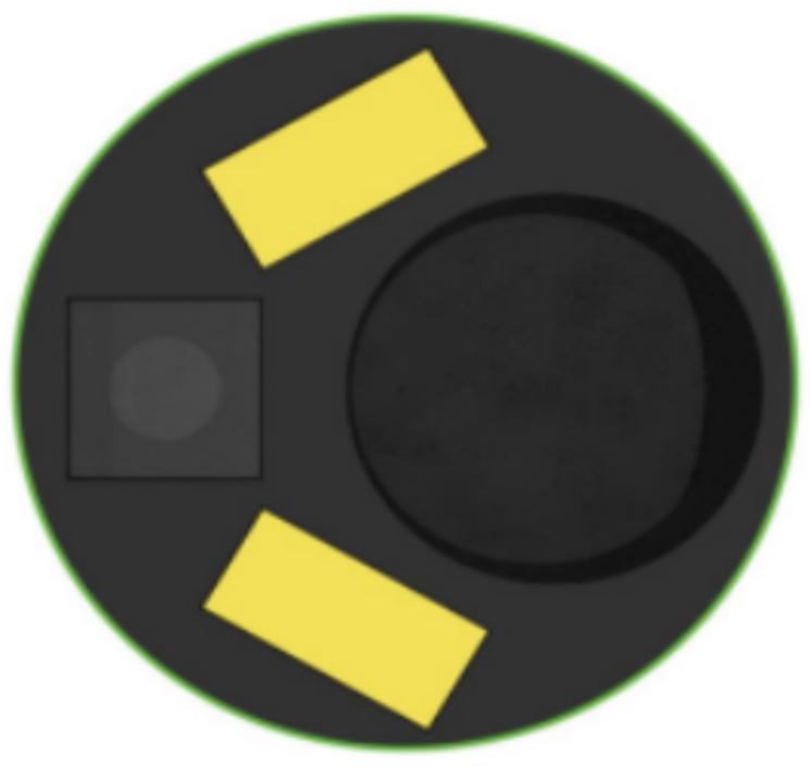
Front Tip
◾ Inner diameter :
Include: Object lens, LED light source.
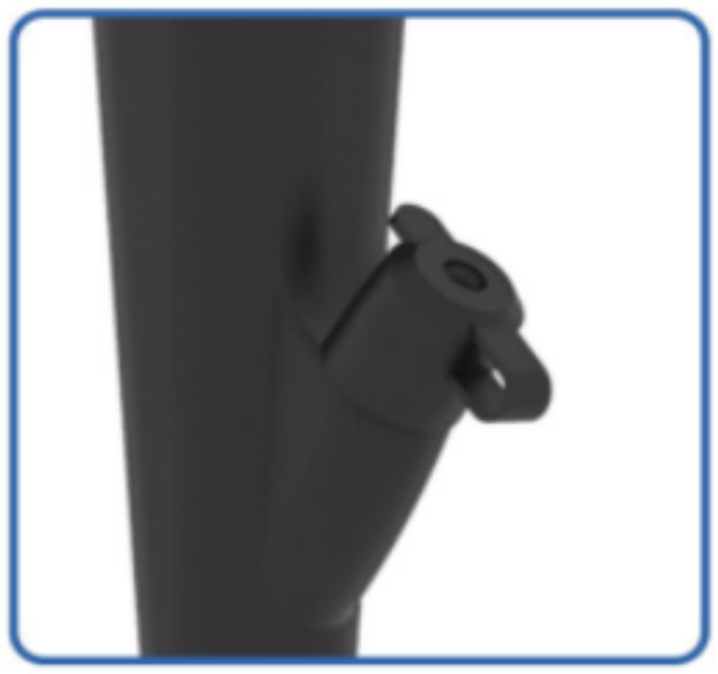
Biopsy channel outlet
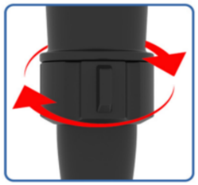
Rotary Adjustment
◾ Rotation can be adjusted by 90° left and right
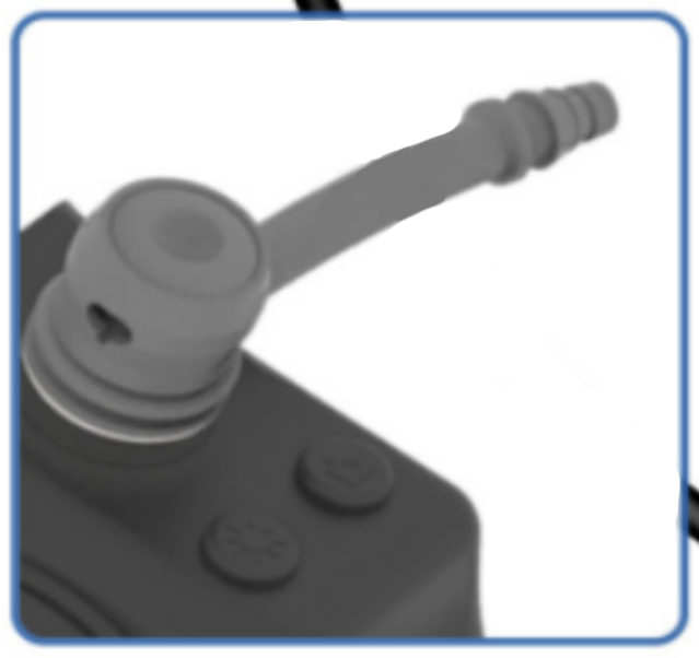
Suction Button
◾ Fast Connection:
Smooth Suction
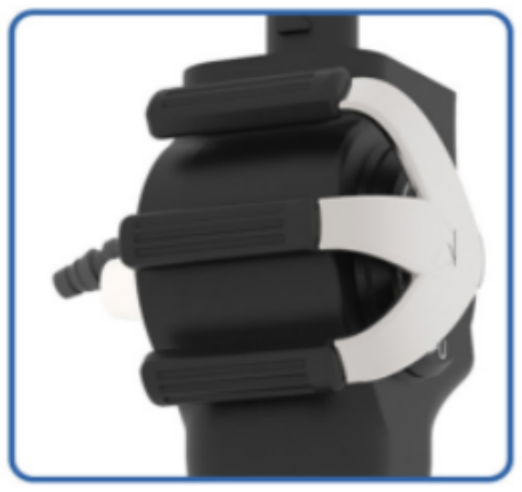
Angulation Control
◾ Flexible angulation control (Up/Down)
◾ Precise location
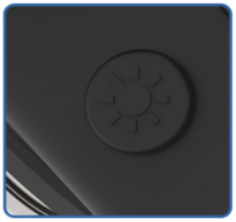
Adjust Brightness
◾ There are 4 gears for adjustment.
 Working Channel
◾ Inner diameter :
3.0mm/2.6mm/2.0mm/1.2mm available

Distal Tip Part
Dual deflections
New CMOS Technology